


EquiCOG
EquiCOG
100 in stock
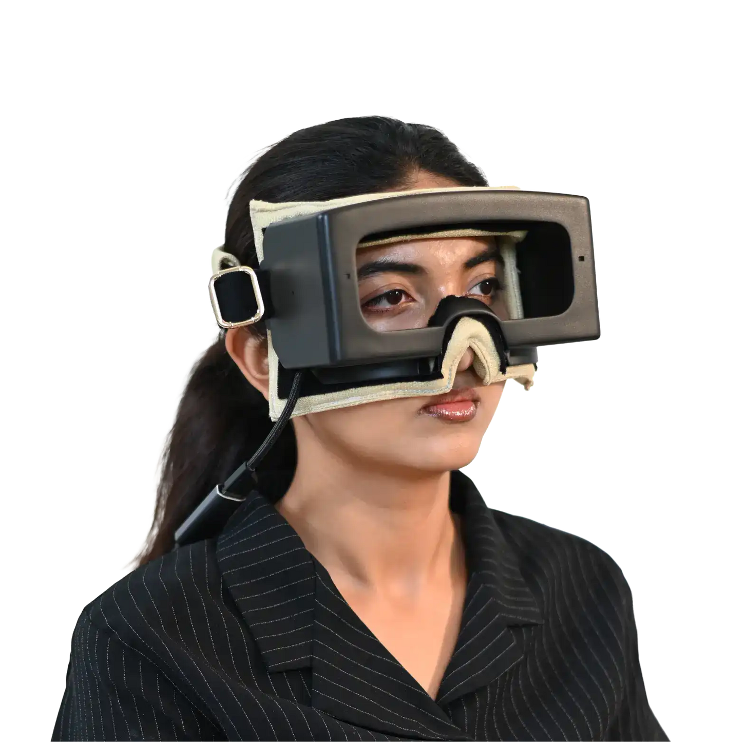
EquiCOG is an advanced videonystagmography (VNG) device that records eye and head movements simultaneously to assess vestibular and ocular motor function.
This VNG device helps diagnose dizziness, vertigo, and balance disorders with high precision using real-time, 3D eye-head tracking.
What's included?
- Goggle with Headband
- Spare Foam Padding
- Calibration Tape (Marked for TV Sizes)
- 5-Meter USB Connecting Cable


Product Features
How EquiCOG works?
- Captures real-time eye movements using dual infrared cameras.
- Measures head motion through built-in inertial sensors for precise tracking.
- Synchronizes head and eye data to analyze vestibulo-ocular coordination.
- Processes signals instantly to display live eye traces and movement graphs.
- Uses automated software algorithms to quantify nystagmus and reflex responses.
- Allows slow-motion review to identify subtle or complex eye movement patterns.
Key features of EquiCOG
- Real-time head tracking along with eye movement.
- High-speed camera records at 120 fps. (100 fps)
- Tracks torsional, horizontal, and vertical eye movements.
- Simultaneous video of head orientation and eye movements.
- Playback in slow motion for precise analysis.
- Generates detailed, easy-to-interpret diagnostic reports with visuals and data.
- Ensures patient comfort with a lightweight, ergonomic goggles design.
- Integrates optional pupillometry and video Frenzel modes for comprehensive evaluation.
Advantages of VNG over ENG
Note: This section is optional. You may choose to omit this section based on the template of the webpage.
VNG has become the preferred method for vestibular testing due to multiple advantages:
- Higher accuracy and sensitivity: VNG’s video camera offers high resolution and stable recording of eye movements, capturing even subtle or torsional nystagmus that ENG (which records only horizontal/vertical via electrodes) would miss. Studies have noted VNG’s superiority in resolution and ability to document torsional eye movements compared to ENG.
- Better patient tolerance: VNG device is relatively quick to fit and not uncomfortable for most patients (similar to wearing ski goggles). In contrast, ENG requires scrubbing skin and attaching electrodes around the eyes, which is time-consuming and can be uncomfortable. VNG also avoids issues like electrode drift or skin impedance problems that can plague ENG recordings.
- Visual confirmation and record: With VNG, clinicians can observe the eye movement on video in real-time and playback important moments in slow motion. This is invaluable for teaching, second opinions, or tracking changes over time. ENG only provides abstract tracings of electrical signals, not an actual video of the eye.
- Torsional nystagmus detection: ENG cannot measure torsional (rotational) movements at all. VNG can at least visually record torsion, and advanced systems can even quantify it. This matters, for example, in diagnosing complex BPPV or central nystagmus that has a torsional component.
- Fewer artifacts: ENG readings can be affected by blinking, lighting, or retinal disorders (since it relies on corneo-retinal potentials). VNG cameras track the pupil directly, so you avoid many such artifacts. However, one limitation: VNG requires the eyes to be open and visible; if a patient’s eyes can’t open well or have heavy ptosis, ENG might be used in that special scenario.
- Efficiency: Overall test time with VNG tends to be shorter since setup is faster (no electrode placement/calibration). Cleanup is also easier (just disinfect the goggles) compared to ENG electrode cleanup. This means better clinical workflow and patient throughput with VNG.
Clinical Uses of EquiCOG
Note: You may choose to use only the headings in the following section. The selection is at the discretion of the designer.
Comprehensive vestibular assessment: EquiCOG is used to evaluate dizziness, vertigo, and balance disorders by recording eye and head movements simultaneously.
Diagnosis of BPPV (Benign Paroxysmal Positional Vertigo): Helps identify the affected semicircular canal and confirms positional nystagmus patterns.
Detection of vestibular neuritis or labyrinthitis: Quantifies unilateral vestibular weakness through spontaneous and caloric-induced nystagmus.
Assessment in Ménière’s disease: Captures episodic nystagmus and evaluates vestibular function between and during attacks.
Evaluation of central vestibular disorders: Differentiates peripheral from central lesions by analyzing abnormal ocular motor findings (e.g., gaze-evoked or vertical nystagmus).
Chronic or complex dizziness evaluation: Offers detailed eye–head correlation to detect subtle abnormalities missed in conventional tests.
Posturography integration: Complements postural stability assessments (e.g., when used alongside EquiPoise) for a full vestibulo-ocular and vestibulo-spinal analysis.
Tracking recovery or rehabilitation: Monitors vestibular compensation and improvement after therapy or surgery.
Educational and clinical training tool: Used in teaching hospitals to visualize real-time vestibular and ocular motor physiology.
Who's a candidate?
- Patients with dizziness or vertigo of unknown cause.
- Individuals with recurrent imbalance or unsteadiness.
- Suspected cases of Benign Paroxysmal Positional Vertigo (BPPV).
- Patients with vestibular neuritis or labyrinthitis.
- Individuals diagnosed with or suspected of Ménière’s disease.
- Patients showing central ocular motor abnormalities.
- Those with chronic or complex dizziness not explained by basic tests.
- Post-head injury or post-ear surgery patients needing vestibular evaluation.
- Elderly or fall-prone individuals with suspected vestibular dysfunction.
- Patients undergoing vestibular rehabilitation to monitor progress.
FAQs
What is VNG?
Videonystagmography (VNG) is a test that uses special goggles with cameras to track your eye movements. It helps doctors check how well your inner ear and brain control balance. The eyes move in certain ways when your head moves, and VNG records that.
What is the clinical relevance of the VNG test?
VNG is the gold standard test for dizziness that helps doctors figure out if vertigo comes from your inner ear or your brain. Healthy balance relies on your inner ear, vision, and body-position sense working together. When the ear’s balance system isn’t working, you get jerky eye movements (nystagmus) and feel dizzy. By recording these eye movements, VNG lets doctors diagnose problems like inner-ear inflammation or brain issues.
What are the advantages of VNG over ENG?
VNG goggles use high-resolution video to catch every eye movement, even tiny twists, that ENG’s electrodes miss. They’re comfortable and quick to put on like ski goggles, avoiding sticky wires and skin preparation. Clinicians can watch and replay real eye videos for clearer diagnosis and teaching, instead of reading electrical blips. With fewer artifacts, faster setup, and easy cleanup, VNG delivers cleaner, more efficient vestibular testing.
What are the key disorders diagnosed by VNG?
VNG (Video Nystagmography) doesn’t diagnose specific diseases, but it spots abnormal eye movements that suggest balance issues. When combined with a person’s symptoms, it can help identify inner ear problems like BPPV (tiny crystals out of place), vestibular infections, Ménière’s disease (with vertigo and hearing loss), or even rare tumors like acoustic neuroma. Each condition shows unique movement patterns during testing. Doctors use these clues to figure out what’s going wrong and how best to treat it.
What is the pathophysiology and mechanisms involved in the vestibular system’s imbalance?
The vestibular system in the inner ear helps us keep balance by sending signals to the brain about head movement and position. When this system is damaged or disrupted due to infection, injury, or other causes, it creates confusion in these signals. This mismatch can make the brain think you're moving when you're not, leading to dizziness, vertigo, or unsteadiness. The brain tries to adapt, but symptoms can vary depending on where the problem is, whether it is inner ear or brain.
What are the types of nystagmus?
Nystagmus is uncontrolled eye movement, and VNG can detect different types based on direction and cause. Horizontal (side-to-side) often signals inner ear issues, while vertical (up or down) or gaze-evoked types may hint at brain problems. Torsional (twisting) movement is common in certain vertigo types like BPPV. Nystagmus may also appear with no trigger (spontaneous) or when changing head position (positional), helping doctors understand where the issue lies.
What is EquiCOG, and how is it different from traditional VNG?
EquiCOG is a next-generation videonystagmography system that not only records high-resolution eye movements but also simultaneously tracks head motion in 3D. Unlike traditional VNG, it includes torsional eye tracking, guided test workflows, and advanced analysis for better diagnostic precision.
How does EquiCOG work?
EquiCOG is a high-tech headset that tracks both eye and head movements in real time using special cameras and sensors. It can capture even tiny, fast eye motions in 3D, including twisting ones most systems miss. By syncing head and eye data, it shows how the eyes react to movement or position changes. This helps doctors accurately detect balance problems even if the patient moves during testing.
Who should be tested using EquiCOG?
Any patient with dizziness, vertigo, imbalance, visual instability, suspected BPPV, Ménière’s, or central vestibular issues. It’s especially helpful in complex or chronic dizziness cases.
How long does a complete EquiCOG test session take?
Depending on the protocol, 20–45 minutes. The streamlined workflow, guided prompts, and quick setup make it significantly faster than traditional systems.
Can EquiCOG differentiate between central and peripheral vertigo?
Yes. Features such as fixation suppression, torsional direction, gaze-evoked nystagmus, and ocular motor test abnormalities help identify central lesions. Combined head and eye data improve localization.
Does EquiCOG support caloric testing?
Yes. It fully supports caloric protocols and provides precise eye movement tracing during thermal stimulation of each ear.
Can EquiCOG be used to diagnose BPPV?
Absolutely. It offers real-time torsional tracking and head angle monitoring during Dix-Hallpike and positional tests, making canal identification more accurate.
What is the advantage of torsional nystagmus tracking?
Most traditional VNGs ignore torsional (rotary) components. EquiCOG quantifies torsion, which is critical for diagnosing BPPV, central causes, and differentiating cupulolithiasis from canalithiasis.
What is Video Frenzel Mode in EquiCOG?
This mode darkens the patient’s vision to remove fixation. The clinician gets a magnified video feed to observe nystagmus more clearly, ideal for spontaneous or subtle positional findings.
Can EquiCOG be used during rotational chair testing?
Yes, EquiCOG goggles can be worn inside compatible rotational chairs, capturing synchronized head and eye movements during rotation.
What specialties can benefit from EquiCOG?
ENT, Neurology, Audiology, Vestibular Therapy, Geriatrics, and Physical Therapy. It is especially effective in interdisciplinary vertigo clinics.
How does EquiCOG provide ROI for clinics?
In-house diagnostics save referrals, increase patient throughput, reduce test time, and enhance diagnostic accuracy—translating to more billed procedures and better outcomes.
Do I need separate devices for vHIT or posturography?
EquiCOG focuses on VNG, but Taevas offers EquiFHit for vHIT and Equipoise for posturography. They are complementary and can be integrated as a suite.
Is prior VNG experience needed to use EquiCOG?
No. The system is designed for ease of use with real-time visual prompts, head angle indicators, and a user-friendly interface. Basic training is sufficient.
What is the frame rate of EquiCOG’s cameras?
EquiCOG uses dual high-resolution infrared cameras recording at 120 frames per second per eye. This ensures accurate capture of high-speed and micro eye movements for detailed velocity and torsional analysis.
Can EquiCOG record torsional eye movements quantitatively?
Yes. EquiCOG uses iris pattern rotation tracking to compute torsional traces, enabling precise angular velocity and direction plotting—useful in BPPV and central disorders.
What type of head tracking sensors are used?
EquiCOG is equipped with a 3-axis IMU (Inertial Measurement Unit) that includes both gyroscope and accelerometer, allowing for real-time tracking of head position, angle, and velocity.
What is the latency of eye-head synchronization?
Eye and head data streams are time-synchronized with millisecond precision. Real-time plotting ensures sub-frame alignment for dynamic vestibulo-ocular reflex (VOR) correlation.
What software platform does EquiCOG run on?
EquiCOG software runs on a Windows-based PC (minimum i5 processor, 8GB RAM recommended). It uses a proprietary Taevas interface for real-time data capture, analysis, and reporting.
Does EquiCOG software generate automatic reports?
Yes. The software provides auto-generated clinical reports that include graphical eye movement traces, torsional velocity plots, cumulative slow-phase velocity, head movement overlays, and snapshot images or embedded video segments.
Is the software compatible with EMR/EHR systems?
Reports are exportable in PDF, making them compatible with most EMR systems. HL7 integration is possible via API for hospitals using structured data ingestion.
Can the software be used in multi-user environments?
Yes. It supports multi-user logins with role-based access (technician, clinician, admin), and maintains session logs for audit trail compliance.
What video resolution is used for eye recording?
Each infrared camera records in at least 640×480 or higher resolution at 120fps. This allows clean, high-contrast pupil tracking even in low light.
Does EquiCOG include pupil detection and blinking compensation?
Yes. The algorithm uses real-time pupil centroid detection, accounts for eyelid interference, and flags blink artifacts in recorded sessions.
Is there a live preview for both eyes during testing?
Yes. The clinician sees dual-eye live video, numerical traces, and head orientation all on a single interface, in real time.
Can data be exported for research?
Yes. Eye and head movement data can be exported in CSV or XML formats for custom research use. Video segments are also downloadable.
Can the device run offline (without internet)?
Yes. All core testing and report generation is offline-capable. Internet is only needed for updates or remote support.
Can EquiCOG be used inside a rotational chair?
Yes. The goggle headset is secure, balanced, and rotational-chair compatible, allowing synchronized data capture during dynamic rotation protocols.
Can EquiCOG be paired with other Taevas devices?
Yes. EquiCOG integrates smoothly with EquiFHit for high-speed head impulse testing, Equipoise for computerized posturography, and Audecom for audiometric evaluations.
What power source does EquiCOG use?
The goggles are USB-powered via a PC or power bank. No external power supply is required for basic operation.
Are software updates included?
Yes. All customers receive lifetime software updates, including bug fixes, UI improvements, and new protocols (subject to license type).
What certifications does EquiCOG have?
EquiCOG is CE-marked for clinical use and built to IEC 60601 safety standards. FDA clearance is in process (if applicable to your market).
How is EquiCOG installed and supported?
The installation is plug-and-play. The Taevas team provides training, calibration support, and software updates. Ongoing remote support is available.

EquiCOG

EquiCOG
EquiCOG